From Science Magazine: “Chemists find a recipe that may have jump-started life on Earth’

AAAS
From Science Magazine

1
New research spells out the simple chemical steps that may have launched the RNA World. Mark Garlick/Science Source

Oct. 18, 2018
Robert F. Service

In the molecular dance that gave birth to life on Earth, RNA appears to be a central player. But the origins of the molecule, which can store genetic information as DNA does and speed chemical reactions as proteins do, remain a mystery. Now, a team of researchers has shown for the first time that a set of simple starting materials, which were likely present on early Earth, can produce all four of RNA’s chemical building blocks.

Those building blocks—cytosine, uracil, adenine, and guanine—have previously been re-created in the lab from other starting materials. In 2009, chemists led by John Sutherland at the University of Cambridge in the United Kingdom devised a set of five compounds likely present on early Earth that could give rise to cytosine and uracil, collectively known as pyrimidines. Then, 2 years ago, researchers led by Thomas Carell, a chemist at Ludwig Maximilian University in Munich, Germany, reported that his team had an equally easy way to form adenine and guanine [Nature], the building blocks known as purines. But the two sets of chemical reactions were different. No one knew how the conditions for making both pairs of building blocks could have occurred in the same place at the same time.

Now, Carell says he may have the answer. On Tuesday, at the Origins of Life Workshop here, he reported that he and his colleagues have come up with a simple set of reactions that could have given rise to all four RNA bases.

Carell’s story starts with only six molecular building blocks—oxygen, nitrogen, methane, ammonia, water, and hydrogen cyanide, all of which would have been present on early Earth. Other research groups had shown that these molecules could react to form somewhat more complex compounds than the ones Carell used.

To make the pyrimidines, Carell started with compounds called cyanoacetylene and hydroxylamine, which react to form compounds called amino-isoxazoles. These, in turn, react with another simple molecule, urea, to form compounds that then react with a sugar called ribose to make one last set of intermediate compounds.

Finally, in the presence of sulfur-containing compounds called thiols and trace amounts of iron or nickel salts, these intermediates transform into the pyrimidines cytosine and uracil. As a bonus, this last reaction is triggered when the metals in the salts harbor extra positive charges, which is precisely what occurs in the final step in a similar molecular cascade that produces the purines, adenine and guanine. Even better, the step that leads to all four nucleotides works in one pot, Carell says, offering for the first time a plausible explanation of how all of RNA’s building blocks could have arisen side by side.

“It looks pretty good to me,” says Steven Benner, a chemist with the Foundation for Applied Molecular Evolution in Alachua, Florida. The process provides a simple way to produce all four bases under conditions consistent with those believed present on early Earth, he says.

The process doesn’t solve all of RNA’s mysteries. For example, another chemical step still needs to “activate” each of RNA’s four building blocks to link them into the long chains that form genetic material and carry out chemical reactions. But making RNA under conditions like those present on early Earth now appears within reach.

See the full article here .


five-ways-keep-your-child-safe-school-shootings

Please help promote STEM in your local schools.

Stem Education Coalition

From PI: Women in STEM-“Celebrating International Women’s Day”

4

Is it not a shame that we need to have a special day to celebrate women when they are so already fantastic and exceptionally brilliant in the physical sciences?

Check out this blog post-

From The Conversation: Women in STEM – “Perish not publish? New study quantifies the lack of female authors in scientific journals”

“”I have done a couple of STEM events, but there have never been this many girls. There are so many here. It is really empowering. Go girls in STEM!” Eama, Grade 12

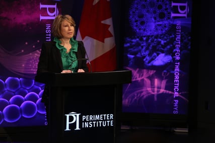
Today’s Inspiring Future Women in Science conference was a success. Mona Nemar, Canada’s Chief Science Advisor, gave opening remarks encouraging the students in attendance to take advantage of the opportunity to learn from the speakers to come.

2
“The days of women being held back or being excluded from science are over. Now, more than ever women are entering, remaining in, and revolutionizing the science fields. Today is a shining example of that.”
-Mona Nemar, Chief Science Advisor, Government of Canada

Mona, read my above post on women getting not published.

The speakers and panelists, who included a chemist, engineer, astronomer, ecologist, and surgeon, talked about the challenges and triumphs that a career in STEM brings. Students were then treated to a speed mentoring session where they were able to ask questions and interact with women from a broad number of STEM careers. Read more about how this conference is inspiring young women here.

3
“This conference showed me there are so many things you can do going into [a career in STEM], so now I feel more inspired, and I feel more confident and not scared to go into science.” Lealan, Age 16

Programs like Perimeter’s “Inspiring Future Women in Science” conference are helping young women see their own potential and reach out for careers in STEM. And more talented female scientists today, means a brighter future tomorrow.

Thank you for being part of the equation.
4

From ICL: “Leukaemia treatment can be made more effective by using a drug for iron overload”

Imperial College London

21 December 2017
Hayley Dunning

1
Healthy bone marrow (yellow) invaded by leukaemia (red), with blood vessels in cyan. Credit: Delfim Duarte

Chemotherapy for one type of leukaemia could be improved by giving patients a drug currently used to treat an unrelated condition, new research shows.

Acute myeloid leukaemia (AML) is an aggressive cancer that stops healthy blood cell production. Chemotherapy is the standard treatment, but improvements are needed as the five-year survival rate in patients older than 60 is only 5-15 per cent.

Now, by studying how leukaemia cells infiltrate bone marrow, where blood cells are created, researchers led by a team from Imperial College London have made a crucial discovery.

Studying mice and human samples, they found that certain areas in the bone marrow support blood stem cells, and when these are overtaken by leukaemia cells, these stem cells are lost and production of healthy blood is significantly reduced. This can cause anaemia, infection, and bleeding in patients, and affects the success of chemotherapy.

Crucially, the team also discovered that a drug already approved to treat a condition known as iron overload can protect these important bone marrow areas and allow blood stem cells to survive. Their results are published today in the journal Cell Stem Cell.

2
Conceptual image from real data of yellow leukaemia invading the bone marrow, dislodging red normal blood cells and destroying cyan blood vessels and blue osteoblasts. Credit: Delfim Duarte

The study’s lead author, Dr Cristina Lo Celso from the Department of Life Sciences at Imperial, said: “Since the drug is already approved for human use for a different condition, we already know that it is safe.

“We still need to test it in the context of leukaemia and chemotherapy, but because it is already in use we can progress to clinical trials much quicker than we could with a brand new drug.”

The researchers are now hoping to team up with clinicians to begin human trials of the drug for AML. Understanding whether this drug is a viable option should take less than five years, as opposed to the 10-15 needed if an entirely new drug is developed.

Protecting blood vessels

The team conducted the study by filming the invasion of leukaemia cells into bone marrow in mice. This approach allowed them see both large overviews and incredible details of the bone marrow, revealing phenomena happening deep inside the bone marrow – a view usually inaccessible to direct observation in patients.

The group discovered that one of the spaces hit particularly hard by leukaemia were special regions of blood vessels where blood stem cells reside. These are the basic blood cells that can become all other types of blood cells, including red and white, generating billions of new cells every day of our life.

For this reason, these special blood vessel regions are vital for producing new healthy blood, and their destruction by leukaemia allows the disease to progress. The loss of these vessels was confirmed in humans by studying patient tissue samples.

3
Blood vessels decreased in bone marrow full of leukaemia (shown with arrows). Credit: Delfim Duarte

To see if they could protect the vessels, the team tested a drug called deferoxamine. The drug is used to treat iron overload, which can happen for example when a person receives multiple blood transfusions.

Deferoxamine has also been used in the treatment of myelodysplasia, a disease related to leukaemia where young blood stem cells do not mature into healthy blood cells. Other researchers who contributed to this project, and are now based at Imperial, Max Plank Munster, and Oxford Kennedy Institute, showed that this drug increases bone marrow vessels in aged mice.

Dr Lo Celso’s group now found that the drug had a protective effect on the blood vessels in AML, allowing the rescue of healthy blood stem cells. Moreover, the enhanced vessels improved the efficiency of chemotherapy.

Delfim Duarte, a physician and PhD student who performed most of the experiments published today, said: “Our work suggests that therapies targeting these blood vessels may improve existing therapeutic regimes for AML, and perhaps other leukaemias too.”

See the full article here .

Please help promote STEM in your local schools.

STEM Icon

Stem Education Coalition

Imperial College London

Imperial College London is a science-based university with an international reputation for excellence in teaching and research. Consistently rated amongst the world’s best universities, Imperial is committed to developing the next generation of researchers, scientists and academics through collaboration across disciplines. Located in the heart of London, Imperial is a multidisciplinary space for education, research, translation and commercialisation, harnessing science and innovation to tackle global challenges.

From EPFL: “The immune cells that help tumors instead of destroying them”

EPFL bloc

École Polytechnique Fédérale de Lausanne EPFL

14.12.17
Nik Papageorgiou

1
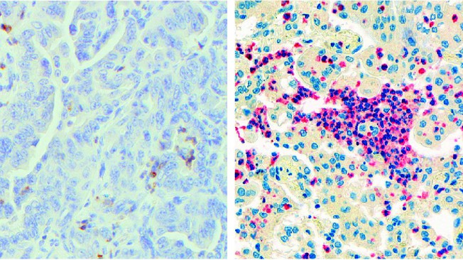
Image: Neutrophils inside lung adenocarcinoma tumors. On the left, neutrophils inside a mouse tumor are stained brown; on the right, neutrophils inside a human tumor are stained red (credit: E. Meylan/EPFL).

EPFL scientists have discovered that neutrophils, a type of immune cell, can actually help lung tumors grow. The work is published in Cell Reports, and has enormous implications for cancer immunotherapy.

Lung cancer is the leading cause of cancer-associated deaths. One of the most promising ways to treat it is by immunotherapy, a strategy that turns the patient’s immune system against the tumor. In the past few years, immunotherapies have been largely based on the degree by which immune cells can infiltrate a tumor, which has become a major predictor of the patient’s overall prognosis.

The problem is that lung tumors adapt to the attacks and find ways to evade them. One of these ways involves a protein called “Programmed death-ligand 1 (PDL1)”, which the tumor cells express on their surface. When immune cells, e.g. T cells, attack the tumor, PDL1 binds a protein on their own surface appropriately named “programmed cell death protein 1”, or PD-1). This interaction triggers an entire cascade of biological reactions in the immune cells that shuts down their attack machinery and renders them harmless to the tumor.

To deal with this, immunotherapeutic regimens often involve drugs that block PD-1, so as to cut off the tumor-evading mechanism. But, unfortunately, this has not been enough. What we need is a fuller understanding of the immune circuits that are active in lung cancer; a knowledge that would allow us to optimize and increase the efficiency of current immunotherapies.

Neutrophils help lung tumors hide

To do this, the lab of Etienne Meylan at EPFL used a mouse model of lung cancer to establish what we call an “immune signature” in lung cancer. The study shows that lung tumors can actually be helped by neutrophils – a type of immune cells that are normally at the first line of attack in infections, allergic reactions, and asthma. In short, neutrophils contribute to disease progression rather than stop it.

The scientists carried out what is known as “neutrophil depletion”, which is a method for studying what happens in a tumor when neutrophil numbers are reduced. By depleting neutrophils in the mice, the researchers were able to deduce what effects they have on a lung tumor when they are actually present.

Surprisingly, depleting neutrophils caused a profound re-modeling of the immune compartment of the lung tumor, with T cells flooding it. This means that neutrophils actually help the tumor hide better from T cells – this is referred to as “immune exclusion”. On the contrary, neutrophil depletion sensitized tumors to anti-PD1 immunotherapies.

“Since neutrophils are important in fighting pathogens, neutrophil depletion is unlikely to be used in the clinic,” says Meylan. “Instead, we must concentrate our efforts to understand exactly how neutrophils promote lung tumor development. This could lead to the identification of drugs that block this specific pro-tumor function of neutrophils.”

Neutrophils help lung tumors grow

The data also showed that the presence of neutrophils leads to changes in the function of the tumor’s blood vessels. The changes trigger hypoxia and cause the tumor cells to produce a protein called “Snail”. This is important because Snail is widely known to help cancer cells resist drugs, as well as promote tumor recurrence and metastasis.

The researchers found that Snail in turn increased the secretion of the protein Cxcl2, augmenting neutrophil infiltration. This creates a positive loop that accelerates the progression of the cancer.

1
In short, the study shows that neutrophils promote tumor progression and can actually hamper the work of immunotherapy in lung cancer. The authors describe this as a “vicious cycle” between neutrophils and Snail that ultimately maintains a tumor microenvironment supporting tumor growth.

“Immunotherapies constitute new treatment options with important clinical success for this devastating disease,” says Etienne Meylan. “But in up to two thirds of patients the lung tumors do not respond. We believe our work offers one explanation for this; finding new ways to break the vicious dialogue between neutrophils and tumor cells might impair tumor growth, and also increase the percentage of patients that benefit from immunotherapy.”

Contributors

Centre Hospitalier Universitaire Vaudois,
University of Lausanne
Swiss Institute of Bioinformatics (VITAL-IT and Bioinformatics Core Facility)
EPFL Flow Cytometry Core Facility
University of Bern

Funding

Swiss National Science Foundation
National Centre of Competence in Research (NCCR) Molecular Oncology,
Swiss Cancer League
Chercher et Trouver Foundation,
ISREC Foundation (“Molecular Life Sciences” grant)
Nuovo Soldati Foundation

See the full article here .

Please help promote STEM in your local schools.

STEM Icon

Stem Education Coalition

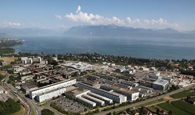
EPFL campus

EPFL is Europe’s most cosmopolitan technical university with students, professors and staff from over 120 nations. A dynamic environment, open to Switzerland and the world, EPFL is centered on its three missions: teaching, research and technology transfer. EPFL works together with an extensive network of partners including other universities and institutes of technology, developing and emerging countries, secondary schools and colleges, industry and economy, political circles and the general public, to bring about real impact for society.

From Rutgers: “Faster, More Accurate Cancer Detection Using Nanoparticles, Rutgers-Led Study Finds”

Rutgers University
Rutgers University

December 11, 2017
Todd B. Bates
848-932-0550
todd.bates@rutgers.edu

Light-emitting nanoprobes can detect cancer early and track the spread of tiny tumors.

1
This illustration shows how human breast cancer cells in a mouse model were “chased” with novel rare earth nanoscale probes injected intravenously. When the subject is illuminated, the probes glow in an infrared range of light that is more sensitive than other optical forms of illumination. In this case, the probes show the spread of cancer cells to adrenal glands and femur (thigh) bones.
Image: Harini Kantamneni and Professor Prabhas Moghe/Rutgers University-New Brunswick.

Using light-emitting nanoparticles, Rutgers University-New Brunswick scientists have invented a highly effective method to detect tiny tumors and track their spread, potentially leading to earlier cancer detection and more precise treatment.

The technology could improve patient cure rates and survival times.

“We’ve always had this dream that we can track the progression of cancer in real time, and that’s what we’ve done here,” said Prabhas V. Moghe, a corresponding author of the study and distinguished professor of biomedical engineering and chemical and biochemical engineering at Rutgers-New Brunswick. “We’ve tracked the disease in its very incipient stages.”

The study, published online Dec.11 in Nature Biomedical Engineering, shows that the new method is better than magnetic resonance imaging (MRI) and other cancer surveillance technologies. The research team included Rutgers’ flagship research institution (Rutgers University-New Brunswick) and its academic health center (Rutgers Biomedical and Health Sciences, or RBHS).

“The Achilles’ heel of surgical management for cancer is the presence of micro metastases. This is also a problem for proper staging or treatment planning. The nanoprobes described in this paper will go a long way to solving these problems,” said Steven K. Libutti, director of Rutgers Cancer Institute of New Jersey. He is senior vice president of oncology services for RWJBarnabas Health and vice chancellor for cancer programs for Rutgers Biomedical and Health Sciences.

The ability to spot early tumors that are starting to spread remains a major challenge in cancer diagnosis and treatment, as most imaging methods fail to detect small cancerous lesions. But the Rutgers study shows that tiny tumors in mice can be detected with the injection of nanoprobes, which are microscopic optical devices, that emit short-wave infrared light as they travel through the bloodstream – even tracking tiny tumors in multiple organs.

The nanoprobes were significantly faster than MRIs at detecting the minute spread of tiny lesions and tumors in the adrenal glands and bones in mice. That would likely translate to detection months earlier in people, potentially resulting in saved lives, said Vidya Ganapathy, a corresponding author and assistant research professor in the Department of Biomedical Engineering.

“Cancer cells can lodge in different niches in the body, and the probe follows the spreading cells wherever they go,” she said. “You can treat the tumors intelligently because now you know the address of the cancer.”

The technology could be used to detect and track the 100-plus types of cancer, and could be available within five years, Moghe said. Real-time surveillance of lesions in multiple organs should lead to more accurate pre- and post-therapy monitoring of cancer.

“You can potentially determine the stage of the cancer and then figure out what’s the right approach for a particular patient,” he said.

In the future, nanoprobes could be used in any surgeries to mark tissues that surgeons want to remove, the researchers said. The probes could also be used to track the effectiveness of immunotherapy, which includes stimulating the immune system to fight cancer cells.

The study includes 16 authors at the School of Engineering (departments of Biomedical Engineering, Chemical and Biochemical Engineering and Materials Science and Engineering) at Rutgers–New Brunswick, the Rutgers Cancer Institute of New Jersey, the Department of Computer Science, and Singapore University of Technology and Design (SUTD).

The study’s lead authors are Harini Kantamneni in the Rutgers Department of Chemical and Biochemical Engineering and Margot Zevon in the Department of Biomedical Engineering. The third corresponding author is Mark C. Pierce, an associate professor in the Department of Biomedical Engineering. The collaboration with Mei-Chee Tan at SUTD was integral to this team effort. Other Rutgers–New Brunswick or RBHS co-authors are Michael J. Donzanti, Shravani R. Barkund, Lucas H. McCabe, Whitney Banach-Petrosky, Laura M. Higgins, Shridar Ganesan, Richard E. Riman and Charles M. Roth.

See the full article here .

Please help promote STEM in your local schools.

STEM Icon

Stem Education Coalition
rutgers-campus

Rutgers, The State University of New Jersey, is a leading national research university and the state’s preeminent, comprehensive public institution of higher education. Rutgers is dedicated to teaching that meets the highest standards of excellence; to conducting research that breaks new ground; and to providing services, solutions, and clinical care that help individuals and the local, national, and global communities where they live.

Founded in 1766, Rutgers teaches across the full educational spectrum: preschool to precollege; undergraduate to graduate; postdoctoral fellowships to residencies; and continuing education for professional and personal advancement.

Rutgers smaller
Please give us back our original beautiful seal which the University stole away from us.
As a ’67 graduate of University college, second in my class, I am proud to be a member of

Alpha Sigma Lamda, National Honor Society of non-tradional students.

From Mapping Cancer Markers at WCG: “New Facility and Expanded Plans for the Mapping Cancer Markers Research Team”

New WCG Logo

WCGLarge

World Community Grid (WCG)

30 Nov 2017
Dr. Igor Jurisica
Krembil Research Institute, University Health Network, Toronto

Summary
The Mapping Cancer Markers researchers recently moved to a new institute, but continues analyzing results, planning for expanded research, and creating new work units throughout the transition. Learn about their plans in this article.

1
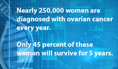
The Mapping Cancer Markers project is currently processing data to identify biomarkers for ovarian cancer, one of the deadliest cancers for women.

After more than 17 years at Ontario Cancer Institute (now Princess Margaret Cancer Centre), we got an opportunity to join Krembil Research Institute (KRI) to work on a more complex approach to chronic diseases, where we moved in mid-November. KRI is still part of the University Health Network in Toronto, but focuses also on arthritis, neuroscience, and vision. The research and translational clinical research interests focus not only on diagnosis and improved treatment, but importantly on prevention, which aligns with my group’s interest over the last few years.

Thus, MCM has not been negatively affected—the physical move was smooth, and our severs stayed in the original server rooms, reducing the risk of any hiccup for World Community Grid work units and results. We will not only continue, but will expand on our research.

New Computational Biology Platform

Importantly, we are in the finalizing stages of paperwork to embark on a newly-funded research by the Ontario Government: The Next Generation Signalling Biology Platform. This will provide us funds to create a software infrastructure for the comprehensive, integrative computational biology analyses workflows, in collaboration with translational research and clinical trials groups. Our partners are Princess Margaret Cancer Centre, Krembil Research Institute, University of Toronto, University of Montreal, BC Cancer Agency, Cancer Clinical Trials Group, Queen’s University, and European Bioinformatics Institute (IMEx Consortium). Besides focusing on cancer, this is our first large osteo-arthritis research project.

Thank you to everyone for your support, and we look forward to providing additional updates as our work progresses.

See the full article here.

Ways to access the blog:
https://sciencesprings.wordpress.com
http://facebook.com/sciencesprings

Please help promote STEM in your local schools.
STEM Icon

Stem Education Coalition

World Community Grid (WCG) brings people together from across the globe to create the largest non-profit computing grid benefiting humanity. It does this by pooling surplus computer processing power. We believe that innovation combined with visionary scientific research and large-scale volunteerism can help make the planet smarter. Our success depends on like-minded individuals – like you.”
WCG projects run on BOINC software from UC Berkeley.
BOINCLarge

BOINC is a leader in the field(s) of Distributed Computing, Grid Computing and Citizen Cyberscience.BOINC is more properly the Berkeley Open Infrastructure for Network Computing.

BOINC WallPaper

CAN ONE PERSON MAKE A DIFFERENCE? YOU BET!!

My BOINC
MyBOINC
“Download and install secure, free software that captures your computer’s spare power when it is on, but idle. You will then be a World Community Grid volunteer. It’s that simple!” You can download the software at either WCG or BOINC.

Please visit the project pages-
Smash Childhood Cancer4

FightAIDS@home Phase II

FAAH Phase II
OpenZika

Rutgers Open Zika

Help Stop TB
WCG Help Stop TB
Outsmart Ebola together

Outsmart Ebola Together

Mapping Cancer Markers
mappingcancermarkers2

Uncovering Genome Mysteries

Say No to Schistosoma

GO Fight Against Malaria

Drug Search for Leishmaniasis

Computing for Clean Water

The Clean Energy Project

Discovering Dengue Drugs – Together

Help Cure Muscular Dystrophy

Help Fight Childhood Cancer

Help Conquer Cancer

Human Proteome Folding

FightAIDS@Home

faah-1-new-screen-saver

faah-1-new

World Community Grid is a social initiative of IBM Corporation
IBM Corporation
ibm

IBM – Smarter Planet
sp

From Yale: “Cellular clean-up can also sweep away forms of cancer”

Yale University bloc

Yale University

November 9, 2017
Bill Hathaway

1
Cells treated (right) or untreated (left) with a PROTAC that degrades the target protein (green). No image credit.

Two new research papers reinforce the benefits of a novel therapy that hijacks the cell’s own protein degradation machinery to destroy cancer cells, Yale researchers report Nov. 9 in the journal Cell Chemical Biology.

The new approach to drug discovery, called Proteolysis Targeting Chimeras or PROTACs, was developed in the laboratory of Craig Crews, the Lewis B. Cullman Professor of Molecular, Cellular, and Developmental Biology, professor of chemistry and pharmacology, as well as executive director of the Yale Center for Molecular Discovery.

The system engages the cell’s own protein degradation machinery to destroy targeted proteins by tagging them for removal. Most drugs are based on the ability of small molecules to bind to and block the function of disease-causing proteins, but some proteins are resistant to such intervention.

“This system will help us change the current small-molecule drug paradigm that fails to target 75% of rogue proteins,” said Crews, scientific founder of Arvinas LLC, the New Haven biotechnology company developing the concept.

The first paper, [Cell Chemical Biology] shows for the first time that PROTAC system can target mutant RTK proteins, which have been linked to several forms of cancer. The second paper [Cell Chemical Biology] proves that the PROTAC system can target rogue proteins with greater specificity than traditional approaches.

Yale’s George M. Burslem and Blake E. Smith are first authors of the first paper. Smith and Yale’s Daniel P. Bondeson are co-first authors of the second paper.

The two papers were primarily funded by the National Institutes of Health. Crews is a shareholder of Arvinas, which also provided researchers to the projects.

See the full article here .

Please help promote STEM in your local schools.

STEM Icon

Stem Education Coalition

Yale University Campus

Yale University comprises three major academic components: Yale College (the undergraduate program), the Graduate School of Arts and Sciences, and the professional schools. In addition, Yale encompasses a wide array of centers and programs, libraries, museums, and administrative support offices. Approximately 11,250 students attend Yale.

From ESA: “Speeding up cancer screening”

ESA Space For Europe Banner

European Space Agency

18 October 2017
No writer credit

1
Radiology scan being examined. ESA.

Delivering breast cancer screening results in a day instead of today’s standard two weeks is being proposed by an ESA incubator start-up company using paperless technology and online image transfers. Screening vans are already on the streets.

“By applying online connectivity to mobile scanning units we have the potential to radically overhaul mobile breast screening in the UK,” notes Viv Barrett of DEOS Consultancy, a start-up from ESA’s business incubator in Harwell, UK.

With one in eight British women developing breast cancer at least once in their lifetime, mobile screening vans are used in the UK to bring the service closer to people, such as offering scanning near supermarkets.

When set up 28 years ago, X-ray film cassettes were physically carried to hospitals for developing. Today, hard drives with digital images are taken by courier, taxi or mammography staff themselves to a hospital for interpretation. In addition to pushing up the cost and adding delay, it is not an efficient use of the medical staff’s time.

2
Breast cancer screening. ESA.

Viv, a qualified mammographer herself with extensive experience of mobile screening, found this unacceptable. Having seen the benefits of working in a connected environment, she founded DEOS Consultancy in 2015 to develop a more efficient scheme.

“When we started four years ago, we used satellite communication technology to develop our system. It enabled us to develop an automatic system and modernise the breast screening units.

“Now we mostly transport images via 3G/4G networks, still fully automatically without involving medical staff. We use satellites if the local mobile networks are too slow.

“We have halved today’s 42 steps, and cut out paper documentation. Our solution is all online. In addition to cutting costs and saving time, it has improved the accuracy and made the work a lot simpler. And it is quicker and much more customer-friendly.”

Currently, bookings are closed several days in advance and the screenings then follow paper diaries printed and delivered to the vans daily, making last-minute changes difficult to handle.

3
DEOS Consultancy has built a demonstration van to present their paperless breast cancer screening system. Copyright DEOS Consultancy.

“Our appointment system is now live so we can handle the breast scanning on the vans better. If a woman turns up on the wrong day, her record can quickly be accessed and if she is eligible she can be screened anyway.

“Having direct access to the patient data, we can add clinical notes directly into the system at the time of screening. It is much safer than today’s practice of adding sticky notes to the paper documents, and occasionally losing them on the way to the hospital.

“Our focus has been on developing a prototype for our technology that could be used in all screening vans.”

4
DEOS Consultancy demonstration van holds a screening room and a separate waiting area. Copyright DEOS Consultancy.

Several have already been fitted with the DEOS online system and are being used, and a demonstration vehicle has been built.

“From these, we can see that images are typically transferred back to the hospital in just 4–10 minutes. That’s impressive because it normally takes at least 24 hours, and sometimes even up to 2 days.

“With our approach, the women could get their results within a day.”

5
The 50th company to graduate from the ESA Business Incubation Centre at Harwell is DEOS Consultancy which is gearing up to overhaul mobile breast screening in the UK. From left: David Osmond (DEOS co-founder) and Viv Barrett (DEOS Co-founder and CEO), with Sue O’Hare (Operations Manager at ESA BIC Harwell), and Anne Green (UK Science and Technology Facilities Council, STFC ). Copyright STFC

“Being hosted at the ESA Business Incubation Centre Harwell puts us in the perfect position to access the specialist technology and expertise we needed to complete our prototype and network with the right audiences and markets,” notes Viv.

“This has been a critical phase in the development of our business, completed well and quickly thanks to the support we have received.”

DEOS became the 50th start-up company to ‘graduate’ from the Harwell centre since it opened in 2011. To date, 61 companies have joined the centre for typically two years, all exploiting space and satellite technology to develop new products and services for terrestrial applications.

The centre is part of the European-wide network of business incubators run by the ESA’s Technology Transfer Programme.

“Turning a brilliant idea into a viable commercial offering is a huge challenge. We help the start-ups to complete this process and become viable businesses,” said Sue O’Hare, Manager of the Harwell incubator.

“DEOS is a good example of how satellite technology can improve a service that could be life-changing for many.”

See the full article here .

Please help promote STEM in your local schools.

STEM Icon

Stem Education Coalition

The European Space Agency (ESA), established in 1975, is an intergovernmental organization dedicated to the exploration of space, currently with 19 member states. Headquartered in Paris, ESA has a staff of more than 2,000. ESA’s space flight program includes human spaceflight, mainly through the participation in the International Space Station program, the launch and operations of unmanned exploration missions to other planets and the Moon, Earth observation, science, telecommunication as well as maintaining a major spaceport, the Guiana Space Centre at Kourou, French Guiana, and designing launch vehicles. ESA science missions are based at ESTEC in Noordwijk, Netherlands, Earth Observation missions at ESRIN in Frascati, Italy, ESA Mission Control (ESOC) is in Darmstadt, Germany, the European Astronaut Centre (EAC) that trains astronauts for future missions is situated in Cologne, Germany, and the European Space Astronomy Centre is located in Villanueva de la Cañada, Spain.

ESA50 Logo large

From Rutgers: “Primary Care Unable to Adequately Care for Cancer Survivors”

Rutgers University
Rutgers University

Oct 6 2017
Carla Cantor
848-932-0555
ccantor@rutgers.edu

1
Seventy-five percent of cancer survivors are seen in primary care practices, but more must be done to meet their health care needs, says a new study. No image credit.

Primary care medicine is currently not able to meet the health care needs of cancer survivors, despite a decade-long effort by the medical establishment to move long-term survivorship care out of the specialists’ realm, according to a new Rutgers study.

The study, published recently in JAMA Internal Medicine, examined 12 advanced primary care practices selected from a national registry of workforce innovators. Not one had a comprehensive survivorship care program in place.

“This is troubling because these are highly innovative practices that have a national reputation,” said study co-author Benjamin Crabtree, a medical anthropologist who is a professor in the Department of Family Medicine and Community Health, Rutgers Robert Wood Johnson Medical School (RWJMS) and a member of the Rutgers Cancer Institute of New Jersey. “As more and more people survive cancer, there will not be enough oncologists to follow these patients and meet their health care needs.”

According to the National Cancer Institute, there are 15.5 million cancer survivors in the United States, a number expected to increase by 31 percent to 20.3 million, by 2026. The vast majority of these patients are seen in primary care practices.

A decade ago the Institute of Medicine released a seminal report, From Cancer Patient to Survivor: Lost in Translation, outlining the need for well-informed primary care survivorship physicians and identifying the components of care. Survivorship care includes checking for cancer re-occurrence, monitoring long-term effects of radiation and chemotherapy treatment and assessing a patient’s psychological well-being.

The researchers, who over two years spent 10 to 12 days observing each of the practices (based in Colorado, Illinois, Maine, New York, Pennsylvania and Washington) and interviewing clinicians and administrators, identified several barriers to integrating survivorship care into primary medicine.

No distinct clinical category for clinicians to identify cancer survivors exists. “There is no diagnosis code for ‘cancer survivor’ that can be entered into the medical record, which is important if you want physicians to pay attention,” Crabtree said.
Electronic medical records (EHR) used in primary care practices have limited capability to record information on patients’ cancer history and clinicians are not provided with actionable recommendations for follow up care.
Medical records sometimes are lost as patients change clinicians over the years, leaving patients to report their cancer histories to their primary care doctors.

In addition to these issues, primary care physicians are concerned about their knowledge gaps in cancer care and the need to monitor changing information in oncology. “There is nothing in the residency curriculum about cancer survivorship,” Crabtree said. “There is also nothing in Continuing Medical Education courses. It’s just not there.”

Only by correcting these deficiencies, can comprehensive cancer survivorship services move to the forefront of primary care, the study states.

“Seventy-five percent of survivors are seen in primary care,” the authors write, “demonstrating a reliance on primary care to address their needs; however, those needs are currently not being met.”

Other contributors to the paper include Ellen B Rubinstein, Ph.D., formerly RWJMS and now with Department of Family Medicine, University of Michigan, Ann Arbor; Shawna V. Hudson, Ph.D., Jenna Howard O’Malley, Ph.D., Heather Sophia Lee, Ph.D., Alicja Bator, MPH (Department of Family and Community Health, RWJMS); William L. Miller, M.D., Lehigh Valley Health network, Allentown, Pennsylvania: and Jennifer Tsui, Ph.D., Rutgers Cancer Institute of New Jersey.

See the full article here .

Follow Rutgers Research here .

Please help promote STEM in your local schools.

STEM Icon

Stem Education Coalition

rutgers-campus

Rutgers, The State University of New Jersey, is a leading national research university and the state’s preeminent, comprehensive public institution of higher education. Rutgers is dedicated to teaching that meets the highest standards of excellence; to conducting research that breaks new ground; and to providing services, solutions, and clinical care that help individuals and the local, national, and global communities where they live.

Founded in 1766, Rutgers teaches across the full educational spectrum: preschool to precollege; undergraduate to graduate; postdoctoral fellowships to residencies; and continuing education for professional and personal advancement.

Rutgers smaller
Please give us back our original beautiful seal which the University stole away from us.
As a ’67 graduate of University college, second in my class, I am proud to be a member of

Alpha Sigma Lamda, National Honor Society of non-tradional students.

From MedicalXpress: “No clear evidence that most new cancer drugs extend or improve life” Don’t read this.

Medicalxpress bloc

MedicalXpress

[This is so sad, it is why I stay away from most cancer research on this blog.]

October 4, 2017

1
Credit: CC0 Public Domain

Even where drugs did show survival gains over existing treatments, these were often marginal, the results [BMJ] show.

Many of the drugs were approved on the basis of indirect (‘surrogate’) measures that do not always reliably predict whether a patient will live longer or feel better, raising serious questions about the current standards of drug regulation.

The researchers, based at King’s College London and the London School of Economics say: “When expensive drugs that lack clinically meaningful benefits are approved and paid for within publicly funded healthcare systems, individual patients can be harmed, important societal resources wasted, and the delivery of equitable and affordable care undermined.”

The research team analysed reports on cancer approvals by the European Medicines Agency (EMA) from 2009 to 2013.

Of 68 cancer indications approved during this period, 57% (39) came onto the market on the basis of a surrogate endpoint and without evidence that they extended survival or improved the quality of patients’ lives.

After a median of 5 years on the market, only an additional 8 drug indications had shown survival or quality of life gains.

Thus, out of 68 cancer indications approved by the EMA, and with a median 5 years follow-up, only 35 (51%) had shown a survival or quality of life gain over existing treatments or placebo. For the remaining 33 (49%), uncertainty remains over whether the drugs extend survival or improve quality of life.

The researchers outline some study limitations which could have affected their results, but say their findings raise the possibility that regulatory evidence standards “are failing to incentivise drug development that best meets the needs of patients, clinicians, and healthcare systems.”

Taken together, these facts paint a sobering picture, says Vinay Prasad, Assistant Professor at Oregon Health & Science University in a linked editorial.

He calls for “rigorous testing against the best standard of care in randomized trials powered to rule in or rule out a clinically meaningful difference in patient centered outcomes in a representative population” and says “the use of uncontrolled study designs or surrogate endpoints should be the exception not the rule.”

He adds: “The expense and toxicity of cancer drugs means we have an obligation to expose patients to treatment only when they can reasonably expect an improvement in survival or quality of life.” These findings suggest “we may be falling far short of this important benchmark.”

This study comes at a time when European governments are starting to seriously challenge the high cost of drugs, says Dr Deborah Cohen, Associate Editor at The BMJ, in an accompanying feature.

She points to examples of methodological problems with trials that EMA has either failed to identify or overlooked, including trial design, conduct, analysis and reporting.

“The fact that so many of the new drugs on the market lack good evidence that they improve patient outcomes puts governments in a difficult position when it comes to deciding which treatments to fund,” she writes. “But regulatory sanctioning of a comparator that lacks robust evidence of efficacy, means the cycle of weak evidence and uncertainty continues.”

In a patient commentary, Emma Robertson says: “It’s clear to me and thousands of other patients like me that our current research and development model has failed.”

Emma is leader of Just Treatment, a patient led campaign with no ties to the pharmaceutical industry, which is calling for a new system that rewards and promotes innovation, so that more effective and accessible cancer medicines are brought within reach.

Editorial: Do cancer drugs improve survival or quality of life? http://www.bmj.com/content/359/bmj.j4528

Patient commentary: the current model has failed, http://www.bmj.com/content/359/bmj.j4568

Feature: Cancer drugs: high price, uncertain value, http://www.bmj.com/content/359/bmj.j4543

See the full article here .

Please help promote STEM in your local schools.

STEM Icon

Stem Education Coalition

Medical Xpress is a web-based medical and health news service that is part of the renowned Science X network. Medical Xpress features the most comprehensive coverage in medical research and health news in the fields of neuroscience, cardiology, cancer, HIV/AIDS, psychology, psychiatry, dentistry, genetics, diseases and conditions, medications and more.