From Weizmann: “How Does Your Microbiome Grow?”

Weizmann Institute of Science logo

Weizmann Institute of Science

01 Sep 2015
No Writer Credit

It is increasingly clear that the thousands of different bacteria living in our intestinal tract – our microbiome– have a major impact on our health. But the details of the microbiome’s effects are still fairly murky. A Weizmann Institute study that recently appeared in Science suggests approaching this topic from a new angle: Assess how fast the various bacteria grow. This approach is already revealing intriguing links between bacterial growth rates and such conditions as type II diabetes and inflammatory bowel disease. The new computational method can illuminate a dynamic process such as growth from a static “snapshot” of a single sample, and thus it may have implications for both diagnostics and new avenues of research.

Tal Korem and David Zeevi, research students in the lab of Prof. Eran Segal of the Computer Science and Applied Mathematics Department, led this research and collaborated with Jotham Suez, a research student in the lab of Dr. Eran Elinav in the Immunology Department, and Dr. Adina Weinberger, a research associate in Segal’s lab. The study began with the advanced genomic sequencing techniques used in many current microbiome studies, which sequence all of the bacterial DNA in a sample. From the short sequences, they construct a picture of the types of bacteria and their relative abundance. But the Weizmann Institute team realized that this sequencing technique held another type of information.

1
Bacterial growth rates computed with the new method (top, average; bottom, for specific species, red represents faster replication) for a human subject that underwent a radical dietary change. Compared are days in which only white boiled rice was consumed (grey area) and days of normal diet (white area). A global change in bacterial growth dynamics was observed between dietary regimens

“The sample’s bacteria are doing what bacteria do best: making copies of their genomes so they can divide,” says Segal. “So most of the bacterial cells contain more than one genome – a genome and a half, for example, or a genome and three quarters.” Since most bacterial strains have pre-programmed “start” and “finish” codes, the team was able to identify the “start” point as the short sequence that was most prevalent in the sample. The least prevalent, at the other end of the genome, was the DNA that gets copied last. The researchers found that analyzing the relative amounts of starting DNA and ending DNA could be translated into the growth rate for each strain of bacteria.

The group tested this formulation experimentally, first in single-strain cultures for which the growth rate could be controlled and observed, then in multiple animal model systems, and finally in the DNA sequences of human microbiomes, in their full complexity.

Their method worked even better than expected: The estimated bacterial growth rates turned out to be nearly identical to observed growth rates. “Now we can finally say something about how the dynamics of our microbiome are associated with a propensity to disease. Microbial growth rate reveals things about our health that cannot be seen with any other analysis method,” says Elinav.

In their examination of human microbiome data, for example, the group found that particular changes in bacterial growth rates are uniquely associated with type II diabetes; others are tied to inflammatory bowel disease. These associations were not observed in the static microbiome “population” studies. Thus the method could be used in the future as a diagnostic tool to detect disease or pathogen infection early on, or to determine the effects of probiotic or antibiotic treatment. In addition, the scientists hope this new understanding of the microbiome will spur further research into the connections between the complex, dynamic ecosystem inside of us and our health.

Also participating in this research were Tali Avnit-Sagi, Maya Pompan-Lotan, Nadav Cohen and Elad Matot in Segal’s lab; Christoph A. Thaiss and Dr. Meirav Pevsner-Fischer in Elinav’s lab; Dr. Ghil Jona and Prof. Alon Harmelin of the Weizmann Institute; Dr. Alexandra Sirota-Madi and Prof. Ramnik Xavier of Harvard Medical School and the Broad Institute; and Prof. Rotem Sorek of the Weizmann Institute.

Dr. Eran Elinav’s research is supported by the Abisch Frenkel Foundation for the Promotion of Life Sciences; the Gurwin Family Fund for Scientific Research; the Leona M. and Harry B. Helmsley Charitable Trust; the Crown Endowment Fund for Immunological Research; the Adelis Foundation; the Rising Tide Foundation; the Vera Rosenberg Schwartz Research Fellow Chair; Yael and Rami Ungar, Israel; John L. and Vera Schwartz, Pacific Palisades, CA; Alan Markovitz, Canada; Leesa Steinberg, Canada; Andrew and Cynthia Adelson, Canada; the estate of Jack Gitlitz; the estate of Lydia Hershkovich; and Mr. and Mrs. Donald L. Schwarz, Sherman Oaks, CA. Dr. Elinav is the incumbent of the Rina Gudinski Career Development Chair.

Prof. Eran Segal’s research is supported by the Adelis Foundation; the Cecil and Hilda Lewis Charitable Trust; the European Research Council; Mr. and Mrs. Donald L. Schwarz, Sherman Oaks, CA; and Leesa Steinberg, Canada.

See the full article here.

Please help promote STEM in your local schools.

STEM Icon

Stem Education Coalition

Weizmann Institute Campus

The Weizmann Institute of Science is one of the world’s leading multidisciplinary research institutions. Hundreds of scientists, laboratory technicians and research students working on its lushly landscaped campus embark daily on fascinating journeys into the unknown, seeking to improve our understanding of nature and our place within it.

Guiding these scientists is the spirit of inquiry so characteristic of the human race. It is this spirit that propelled humans upward along the evolutionary ladder, helping them reach their utmost heights. It prompted humankind to pursue agriculture, learn to build lodgings, invent writing, harness electricity to power emerging technologies, observe distant galaxies, design drugs to combat various diseases, develop new materials and decipher the genetic code embedded in all the plants and animals on Earth.

The quest to maintain this increasing momentum compels Weizmann Institute scientists to seek out places that have not yet been reached by the human mind. What awaits us in these places? No one has the answer to this question. But one thing is certain – the journey fired by curiosity will lead onward to a better future.

From Cornell: “Antibody-making bacteria promise drug development”

Cornell Bloc

Cornell University

August 31, 2015
Anne Ju

Monoclonal antibodies, proteins that bind to and destroy foreign invaders in our bodies, routinely are used as therapeutic agents to fight a wide range of maladies including breast cancer, leukemia, asthma, arthritis, psoriasis, Crohn’s disease and transplant rejection. Humira, a treatment for arthritis and Crohn’s disease, was among the first lab-engineered antibody drugs.

1
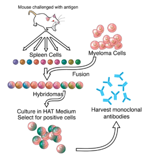
A general representation of the method used to produce monoclonal antibodies

Typically, monoclonal antibodies are manufactured in animal cell lines, such as Chinese hamster ovary (CHO) cells, with long development times that can drive up cost. A team of Cornell chemical engineers and New England Biolabs scientists have devised a shortcut. They’ve done it using an engineered E. coli bacterium that carries machinery for human antibody production and can churn out complex proteins, including many of today’s blockbuster, life-saving antibody drugs, in as little as a week.

A Nature Communications paper published Aug. 27 details the feat, led by co-senior author Matthew DeLisa, the William L. Lewis Professor of Engineering, and first author Michael-Paul Robinson, a graduate student in the field of chemical engineering. They worked with a team led by co-senior author Mehmet Berkmen, a staff scientist at New England Biolabs.

The work built on a previously commercialized E. coli strain invented by Berkmen, called “SHuffle,” which could make shorter, simpler proteins such as antibody fragments that had less therapeutic value than their full-sized, monoclonal antibody counterparts. Now, the researchers report producing full-length antibodies using the specially engineered SHuffle bacterium, including ones that fight the avian flu virus, the anthrax pathogen Bacillus anthracis, and a replica of the therapeutic antibody Herceptin that is used to treat breast cancer.

“We can engineer new antibodies in SHuffle almost as quickly as our bodies can. Customizing an antibody requires only simple edits to the bacterium’s DNA, which opens up a low-effort way to prototype new ideas for future therapeutics,” Berkmen said.

The SHuffle bacterium harbors genetic modifications that allow it, unlike other bacteria, to assemble antibodies and other human proteins into their natural, functional shape. A unique aspect of the method is the “all-in-one-pot” manner in which the large, complicated antibody molecules are assembled, taking place exclusively in the cytoplasmic compartment of the bacterium.

This method effectively bypasses some of the key bottlenecks in the multi-compartment biosynthesis inherent to such production hosts as CHO cells. Preliminary experiments indicate the SHuffle-made antibodies could be recognized by the human immune system as robustly as the originals.

“We think this is going to be a very powerful way of biomanufacturing existing antibodies, or even developing entirely new ones from scratch, that is much faster than current methods,” DeLisa said.

While immunotherapeutics invented in bacteria may one day become useful medicines, other uses may abound.

“Many diagnostic tests, such as those performed on tumor biopsies, depend on finely-tuned antibodies,” DeLisa said. “Scientists also depend upon antibodies to make the molecular mechanics of living organisms visible, but sometimes they lack antibodies that work well enough for their experiments.”

The paper is titled “Efficient expression of full-length antibodies in the cytoplasm of engineered bacteria,” and the work was supported by the National Institutes of Health, the Ford Foundation and the National Science Foundation.

See the full article here.

Please help promote STEM in your local schools.

STEM Icon

Stem Education Coalition
Once called “the first American university” by educational historian Frederick Rudolph, Cornell University represents a distinctive mix of eminent scholarship and democratic ideals. Adding practical subjects to the classics and admitting qualified students regardless of nationality, race, social circumstance, gender, or religion was quite a departure when Cornell was founded in 1865.

Today’s Cornell reflects this heritage of egalitarian excellence. It is home to the nation’s first colleges devoted to hotel administration, industrial and labor relations, and veterinary medicine. Both a private university and the land-grant institution of New York State, Cornell University is the most educationally diverse member of the Ivy League.

On the Ithaca campus alone nearly 20,000 students representing every state and 120 countries choose from among 4,000 courses in 11 undergraduate, graduate, and professional schools. Many undergraduates participate in a wide range of interdisciplinary programs, play meaningful roles in original research, and study in Cornell programs in Washington, New York City, and the world over.

From COSMOS: “Fighting superbugs with supercomputers”

Cosmos Magazine bloc

COSMOS

31 Aug 2015
Viviane Richter

1
Doctors need new ways to attack the antibiotic-resistant bacterium MRSA, shown here growing on a blood agar plate.Credit: By R Parulan Jr./getty images

We’re losing the arms race against superbugs. Now with the aid of a supercomputer, Alan Grossfield at the University of Rochester is refining a new battlefield strategy. Instead of attacking their proteins, which bacteria can disguise, the new weapons attack the membrane which is much harder to hide. Grossfield and his team’s findings were published in Biophysical Journal in August.

In this arms race “the cell membrane is like the final frontier”, says University of Queensland microbiologist Matt Cooper.

Last year, the UK Review on Antimicrobial Resistance estimated that antibiotic resistant bacteria account for at least 700,000 deaths each year and could grow to 10 million worldwide by 2050. If we want to avoid entering a post-antibiotic era we need new armaments.

Most antibiotics are designed to latch on to and deactivate a single protein target in the cell. Penicillin, for example, blocks an enzyme bacteria need to hold their cell wall together. However, bacteria can rapidly mutate these protein targets making them unrecognisable to the antibiotics.

But bacteria are far less able to mutate the structure of their membranes; their basic life chemistry relies on it. So drugs that attack the bacterial membrane should be harder to beat.

Tree frogs discovered this trick long ago. Their skin contains a host of antimicrobial and antifungal defences – including a group called lipopeptides that slice bacterial membranes, making them leaky. Medicinal chemists are now developing lipopeptides of their own, to be used as antimicrobial drugs. The first, daptomycin, is the only new antibiotic to be approved by the US Food and Drug Administration in the past 15 years.

Researchers hope to make other lipopeptide drugs that are even more potent that daptomycin. The problem is that human cells also have membranes – so when designing membrane-slicing drugs, it’s important that bacteria remain their sole target. Grossfield looked more closely at one lipopeptide drug in development, already shown to clear bacterial infections in mice, in order to understand how the drug worked.

Antimicrobial lipopeptides clump together in roughly spherical clusters known as micelles. They float through the bloodstream with their weapons hidden – like a Swiss army knife with all its blades folded away. Only when a clump reaches a target do the blades flip out to pierce the membrane.

With the help of a supercomputer, Grossfield’s team simulated how the drug responded when stuck to a bacterial and mammalian membrane. This drug’s action takes less than 500th of a second, but the simulations took an entire year of number crunching.

It turns out the drug has a slight positive charge. Luckily mammalian membranes are neutral, so the drug doesn’t stick. But bacterial membranes are negatively charged. Once stuck, the drug’s “blades” quickly flick out and slice into the membrane. The team found the drug stabbed bacterial membranes 50 orders of magnitude faster than mammalian membranes. “This was really cool,” Grossfield says.

In this computer simulation, the lipopeptide cluster (green and yellow) sticks to the bacterium’s surface (blue), and then all of a sudden (12 seconds through the video) slices its way into the bacterium’s membrane. Credit: Dejun Lin, University of Rochester Medical Centre

He also found there’s a sweet spot to the drug’s blade length. If they’re too long, they tend to get jammed in the clump. Too short, and they don’t inflict enough damage to kill the bacterium.

He hopes his work will help chemists design better lipopeptides in the future: “I hope I can explain to the medicinal chemists what drug properties they should think about.”

The new drugs will buy us more time, but even this strategy is not likely to last. Some bacteria have already found a defence against daptomycin, first membrane-stabbing drug, which was rolled out in 2003.

Cooper believes over-prescription of antibiotics is the biggest contributor to resistance. But “attacking the membrane gives us more time”, he says. “We want to find drugs that give us a couple of decades.”

See the full article here.

Please help promote STEM in your local schools.

STEM Icon

Stem Education Coalition

From Rockefeller: “Atomic view of cellular pump reveals how bacteria send out proteins”

Rockefeller U bloc

Rockefeller University

July 22, 2015
Wynne Parry | 212-327-7789

1
A watery passage: The pump, a single-molecule machine, (yellow coils) carries proteins through the cell membrane (pink and dark blue). Within the pump, the researchers found a strikingly large water-filled channel (light blue), a natural environment for hydrophilic proteins. No image credit

Bacteria have plenty of things to send out into world beyond their own boundaries: coordinating signals to other members of their species, poisons for their enemies, and devious instructions to manipulate host cells they have infected. Before any of this can occur, however, they must first get the shipments past their own cell membranes, and many bacteria have evolved specialized structures and systems for launching the proteins that do these jobs.

Researchers at The Rockefeller University have determined the structure of a simple but previously unexamined pump that controls the passage of proteins through a bacterial cell membrane, an achievement that offers new insight into the mechanics that allow bacteria to manipulate their environments. The results were published in Nature on July 23.

“This pump, called PCAT for peptidase-containing ATP-binding cassette transporter, is composed of a single protein, a sort of all-in-one machine capable of recognizing its cargo, processing it, then burning chemical fuel to pump that cargo out of the cell,” says study author Jue Chen, William E. Ford Professor and head of the Laboratory of Membrane Biology and Biophysics. “This new atomic-level structure explains for the first time the links between these three functions.”

Of the many types of molecules cells need to move into and out of their membranes, proteins are the largest. PCATs specialize in pumping proteins out of the cell, and, because they are single-molecule machines that work alone, or with two partner proteins in some bacteria, they are the simplest such systems.

Each PCAT molecule has three domains, each in duplicate: one recognizes the cargo by a tag it carries, and cuts off that tag; another binds to and burns ATP, a molecule that contains energy stored within its atomic bonds; and the third forms a channel that spans the cells membrane. Previous work had examined the structure of the first two domains, but the structure of the third, had remained a mystery, along with the details of how the components function together.

“At this point, we have no idea how many PCATs exist, although we expect they are numerous, because each specializes in a specific type of cargo. For this study, we focused on one we called PCAT1, which transports a small protein of unknown function,” says first author David Yin-wei Lin, a postdoc in the lab. “To get a sense of how PCAT1 changes shape when powered by energy from ATP, we examined the structure in two states, both with and without ATP.”

The team, which also included Shuo Huang, a research technician who is now a graduate student at Georgia Institute of Technology, purified and crystalized the PCAT1 protein from the heat-loving bacterium Clostridium thermocellum. To determine the structure of the crystals, they used a technique called X-ray diffraction analysis, in which a pattern produced by X-rays bounced off the crystallized protein can be used to infer the structure of the molecule.

The first structure, determined without ATP, revealed a striking feature: a large, water-filled central channel, a natural environment for a water-loving, or hydrophilic, protein. Two side openings into this channel were guarded by the cargo-recognizing domain, acting as a sort of ticket taker. Sites on this domain would recognize and clip off the cargo’s tag, before ushering the protein into the channel.

When ATP is present, they found that the side entrances close, freeing the cargo-recognizing domain to move from its station outside of them. In addition, the ATP-binding domains at the bottom of the channel inside the cell come together. The researchers also saw the water channel shrink, leading them to hypothesize that energy from ATP allows PCAT1 to change conformation in such a way that it pushes its cargo out. This suggests that PCAT1 uses a strategy commonly seen in transport proteins known as alternate access, in which one end of the channel is open while the other closes. However, they qualify that PCATs that transport much larger proteins may function differently.

“By visualizing the structure of this pump, we have been able to determine the details of a transport pathway that, in its simplicity, is fundamentally different from the more complex systems that have been closely studied before. This new information adds to the understanding of how cells send out proteins in order to interact with their environment,” Chen says.

See the full article here.

Please help promote STEM in your local schools.

STEM Icon

Stem Education Coalition

Rockefeller U Campus

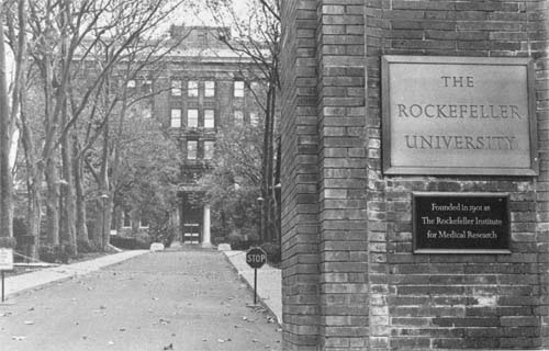
The Rockefeller University is a world-renowned center for research and graduate education in the biomedical sciences, chemistry, bioinformatics and physics. The university’s 76 laboratories conduct both clinical and basic research and study a diverse range of biological and biomedical problems with the mission of improving the understanding of life for the benefit of humanity.

Founded in 1901 by John D. Rockefeller, the Rockefeller Institute for Medical Research was the country’s first institution devoted exclusively to biomedical research. The Rockefeller University Hospital was founded in 1910 as the first hospital devoted exclusively to clinical research. In the 1950s, the institute expanded its mission to include graduate education and began training new generations of scientists to become research leaders around the world. In 1965, it was renamed The Rockefeller University.

From LBL: “First Detailed Microscopy Evidence of Bacteria at the Lower Size Limit of Life”

Berkeley Logo

Berkeley Lab

February 27, 2015
Dan Krotz

1
This cryo-electron tomography image reveals the internal structure of an ultra-small bacteria cell like never before. The cell has a very dense interior compartment and a complex cell wall. The darker spots at each end of the cell are most likely ribosomes. The image was obtained from a 3-D reconstruction. The scale bar is 100 nanometers. (Credit: Berkeley Lab)

Scientists have captured the first detailed microscopy images of ultra-small bacteria that are believed to be about as small as life can get. The research was led by scientists from the U.S. Department of Energy’s Lawrence Berkeley National Laboratory and the University of California, Berkeley. The existence of ultra-small bacteria has been debated for two decades, but there hasn’t been a comprehensive electron microscopy and DNA-based description of the microbes until now.

The cells have an average volume of 0.009 cubic microns (one micron is one millionth of a meter). About 150 of these bacteria could fit inside an Escherichia coli cell and more than 150,000 cells could fit onto the tip of a human hair.

The scientists report their findings Friday, Feb. 27, in the journal Nature Communications.

The diverse bacteria were found in groundwater and are thought to be quite common. They’re also quite odd, which isn’t a surprise given the cells are close to and in some cases smaller than several estimates for the lower size limit of life. This is the smallest a cell can be and still accommodate enough material to sustain life. The bacterial cells have densely packed spirals that are probably DNA, a very small number of ribosomes, hair-like appendages, and a stripped-down metabolism that likely requires them to rely on other bacteria for many of life’s necessities.

The bacteria are from three microbial phyla that are poorly understood. Learning more about the organisms from these phyla could shed light on the role of microbes in the planet’s climate, our food and water supply, and other key processes.

2
A lifeline to other cells? Cryo-transmission electron microscopy captured numerous hairlike appendages radiating from the surface of this ultra-small bacteria cell. The scientists theorize the pili-like structures enable the cell to connect with other microbes and obtain life-giving resources. The scale bar is 100 nanometers. (Credit: Berkeley Lab)

“These newly described ultra-small bacteria are an example of a subset of the microbial life on earth that we know almost nothing about,” says Jill Banfield, a Senior Faculty Scientist in Berkeley Lab’s Earth Sciences Division and a UC Berkeley professor in the departments of Earth and Planetary Science and Environmental Science, Policy and Management.

“They’re enigmatic. These bacteria are detected in many environments and they probably play important roles in microbial communities and ecosystems. But we don’t yet fully understand what these ultra-small bacteria do,” says Banfield.

Banfield is a co-corresponding author of the Nature Communications paper with Birgit Luef, a former postdoctoral researcher in Banfield’s group who is now at the Norwegian University of Science and Technology, Trondheim.

“There isn’t a consensus over how small a free-living organism can be, and what the space optimization strategies may be for a cell at the lower size limit for life. Our research is a significant step in characterizing the size, shape, and internal structure of ultra-small cells,” says Luef.

The scientists set out to study bacteria from phyla that lack cultivated representatives. Some of these bacteria have very small genomes, so the scientists surmised the bacteria themselves might also be very small.

To concentrate these cells in a sample, they filtered groundwater collected at Rifle, Colorado through successively smaller filters, down to 0.2 microns, which is the size used to sterilize water. The resulting samples were anything but sterile. They were enriched with incredibly tiny microbes, which were flash frozen to -272 degrees Celsius in a first-of-its-kind portable version of a device called a cryo plunger. This ensured the microbes weren’t damaged in their journey from the field to the lab.

The frozen samples were transported to Berkeley Lab, where Luef, with the help of Luis Comolli of Berkeley Lab’s Life Sciences Division, characterized the cells’ size and internal structure using 2-D and 3-D cryogenic transmission electron microscopy. The images also revealed dividing cells, indicating the bacteria were healthy and not starved to an abnormally small size.

The bacteria’s genomes were sequenced at the Joint Genome Institute, a DOE Office of Science User Facility located in Walnut Creek, California, under the guidance of Susannah Tringe. The genomes were about one million base pairs in length. In addition, metagenomic and other DNA-based analyses of the samples were conducted at UC Berkeley, which found a diverse range of bacteria from WWE3, OP11, and OD1 phyla.

This combination of innovative fieldwork and state-of-the-art microscopy and genomic analysis yielded the most complete description of ultra-small bacteria to date.

Among their findings: Some of the bacteria have thread-like appendages, called pili, which could serve as “life support” connections to other microbes. The genomic data indicates the bacteria lack many basic functions, so they likely rely on a community of microbes for critical resources.

The scientists also discovered just how much there is yet to learn about ultra-small life.

“We don’t know the function of half the genes we found in the organisms from these three phyla,” says Banfield.

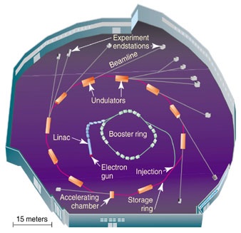
The scientists also used the Advanced Light Source, a DOE Office of Science User Facility located at Berkeley Lab, where Hoi-Ying Holman of the Earth Sciences Division helped determine the majority of the cells in the samples were bacteria, not Archaea.

LBL Advanced Light Source
LBL ALS interior
LBL/ALS

The research is a significant contribution to what’s known about ultra-small organisms. Recently, scientists estimated the cell volume of a marine bacterium at 0.013 cubic microns, but they used a technique that didn’t directly measure the cell diameter. There are also prior electron microscopy images of a lineage of Archaea with cell volumes as small as 0.009 cubic microns, similar to these bacteria, including results from some of the same researchers. Together, the findings highlight the existence of small cells with unusual and fairly restricted metabolic capacities from two of the three major branches of the tree of life.

The research was supported by the Department of Energy’s Office of Science.

See the full article here.

Please help promote STEM in your local schools.

STEM Icon

Stem Education Coalition

A U.S. Department of Energy National Laboratory Operated by the University of California

University of California Seal

DOE Seal

From NOVA: “This Bacterium Can Survive on Electricity Alone”

PBS NOVA

NOVA

21 Jul 2014
Allison Eck

Scientists are hoping that a large battery in a South Dakotan gold mine could lure curious forms of bacteria that may help us understand what powers life as we know it.

That’s because scientists have begun to discover bacteria that live and thrive on electricity alone. Rather than mediating electrons through third-party materials (such as sugar or oxygen) like most organisms do, these bacteria transmit them unaccompanied by anything else. Understanding how these interactions work could give us a glimpse of the kind of life that might exist on other planets.

bact
Geobacter sulfurreducens breathes by transferring electrons to iron oxides found in soil.

Here’s Catherine Brahic, writing for New Scientist:

Unlike any other living thing on Earth, electric bacteria use energy in its purest form—naked electricity in the shape of electrons harvested from rocks and metals. We already knew about two types, Shewanella and Geobacter. Now, biologists are showing that they can entice many more out of rocks and marine mud by tempting them with a bit of electrical juice. Experiments growing bacteria on battery electrodes demonstrate that these novel, mind-boggling forms of life are essentially eating and excreting electricity.

And scientists have found more than just a few new examples. Annette Rowe, a doctoral student at the University of Southern California, Los Angeles, has identified up to eight specimens demonstrating this behavior. That suggests to her advisor, Kenneth Nealson, that there could be a whole slew of organisms involved in direct extraction of electrons.

While the immediate applications are obvious—for example, better biomachines (or self-powered devices) for human use—the findings could also tell us what life’s “bare minimum” is. In other words, at what amount of energy does life begin? And is it possible to create a bacterium that, through electric means, cannot be destroyed?

Brahic again:

For that we need the next stage of experiments, says Yuri Gorby, a microbiologist at the Rensselaer Polytechnic Institute in Troy, New York: bacteria should be grown not on a single electrode but between two. These bacteria would effectively eat electrons from one electrode, use them as a source of energy, and discard them on to the other electrode.

Other-worldly expeditions to mines or deep-sea caves could help us find more examples of organisms that interact with their environments this way, thereby bringing us closer to answering some of these questions.

See the full article here.

NOVA is the highest rated science series on television and the most watched documentary series on public television. It is also one of television’s most acclaimed series, having won every major television award, most of them many times over.

ScienceSprings relies on technology from

MAINGEAR computers

Lenovo
Lenovo

Dell
Dell